


PAST CLASS The Exquisite Art of Human Anatomy: History and Techniques: A Live, Online Class with Medical Illustrator Marie Dauenheimer, MA, CMI, Starting August 30
Tuesdays, August 30 – October 4
Time: 6:00 pm - 8:00 pm New York EST
Admission: $155 Patreon Members / $165 Regular Admission
PLEASE NOTE: All classes will also be recorded and archived for students who cannot make that time
Join medical illustrator Marie Dauenheimer--medical editor of Anatomica: The Exquisite and Unsettling Art of Human Anatomy—for a deep dive into centuries of anatomical art. Each session of this six week class will focus on specific artists and anatomists who worked together to change the way we view and think about the human body.
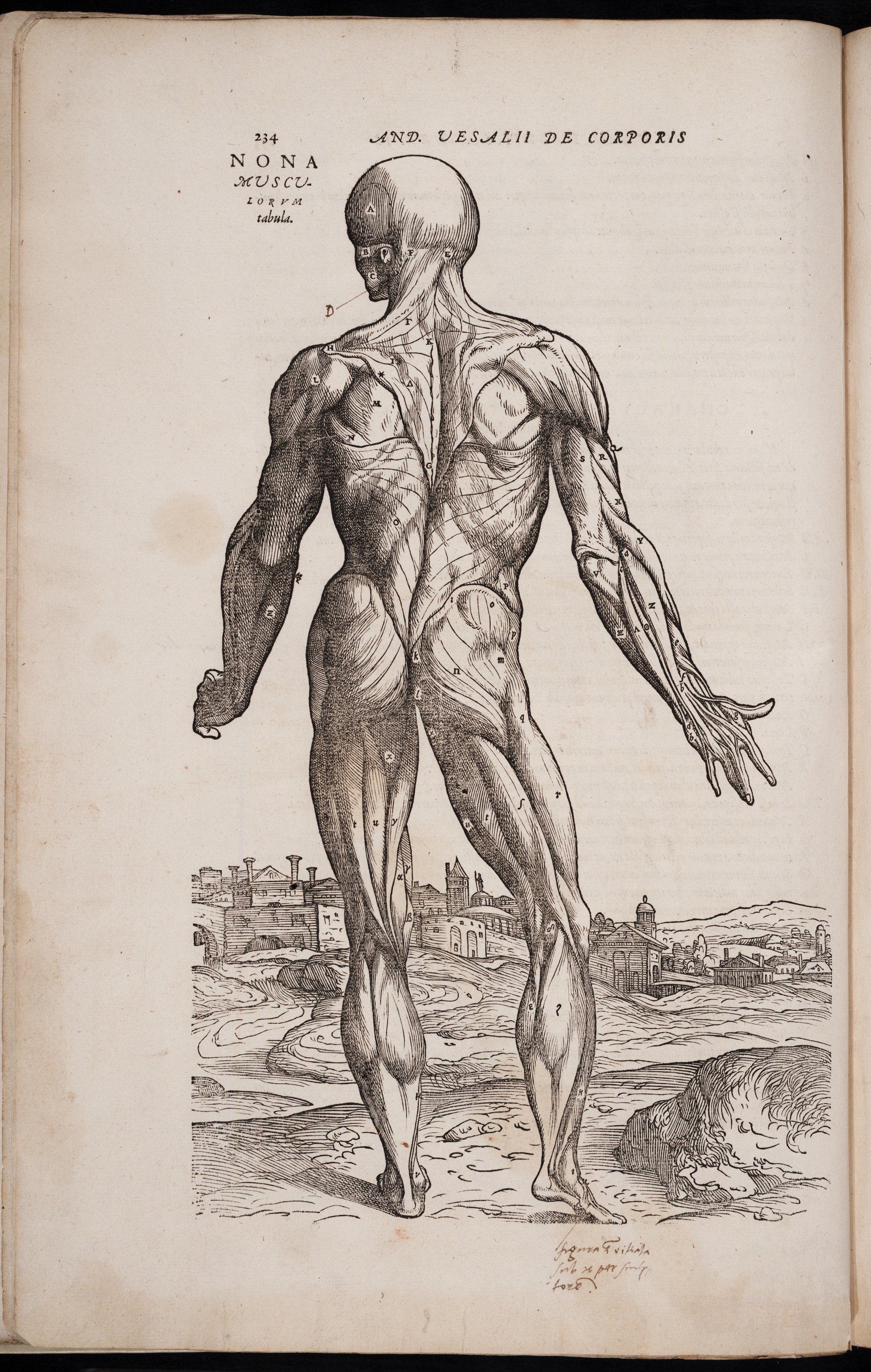
The class will begin with a deep dive into the first illustrated anatomy book, Vesalius’ De Humani Corporis Fabrica (1543), which heralded the birth of modern anatomy, transforming the field. We will learn about the pioneering anatomist Andreas Vesalius, whose knowledge came from his own dissections of cadavers, and Jan Van Calcar, a pupil of the great painter Titian, who is thought to have rendered the evocative woodcuts that illustrate the text.
Next, we’ll look at the macabre skeletal fetal tableaux created by anatomist and physician Dr. Frederik Ruysch. Once on display in a home museum eventually purchased by Tsar Peter the Great, they were beautifully rendered by artist Cornelius Huyberts in Ruysch’s publication Thesuarus Anatomicus.
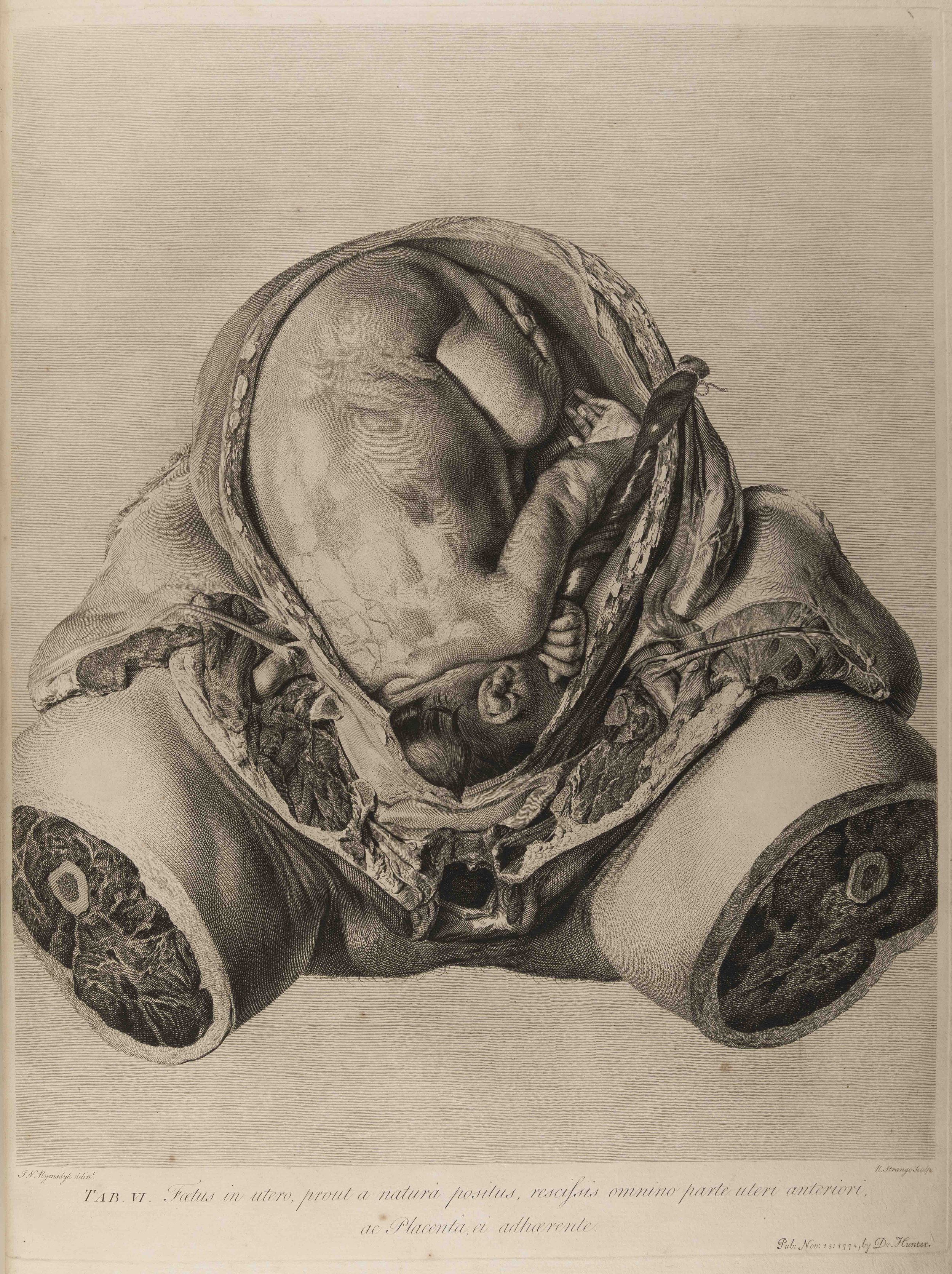
Other classes will take a investigate the revolutionary studies of the gravid (or pregnant) uterus conducted by anatomist William Hunter and artist Jan Van Rymsdyk, resulting in the publication of their magnum opus The Anatomy of the Gravid Uterus (1774); the elegant and idealized “homo perfectus” engravings created by Jan Wanderlaar and Siegfried Bernard Albinus for their tour de force anatomical atlas Tabulae sceleti et musculorum corporis humani of 1749; and the otherworldly, gorgeously colored mezzotints of 18th century artist and anatomist Jacques-Fabien Gautier d'Agoty, creator of the iconc “flayed angel” (see top image) among other memorable works.
Each richly illustrated lecture will explore the fascinating history of anatomical art in richly illustrated lectures focusing on specific artists and anatomists. Each session will end with an in-class art project designed to enhance the understanding of the work. The instructor—an experienced medical illustrator—will guide students in the use of traditional techniques including as carbon dust, colored pencils, pen & ink, and collage.
Suggested materials list will be sent to students before class begins.
Week One: General introduction to the history of anatomical art as outlined in Anatomica. (August 30, 2022)
General drawing assignment, and introduction to materials.
Week Two: Andreas Vesalius and Jan Van Calcar, De Humani Corporis Fabrica (1543) (September 6, 2022)
Lecture: Explore the rich history of the making of Vesalius’s masterpiece De Fabrica. This remarkable book was the first to use detailed and highly accurate anatomical illustrations credited to artist Jan Van Calcar.
Technique: Using pen and ink reinvent and explore the richly textured wood engravings in Da Fabrica.
Week Three: Dr. Frederik Ruysch, Cornelius Huyberts, and the fetal tableaux (17th century) (September 13, 2022)
Lecture: Focus on Ruysch and his amazing tableaux of fetal skeletons.
Technique: Using collage as well as traditional drawing materials create a Ruysch like tableau.
Week Four: 18th century—Jan Van Rymsdyk, William Hunter, the Gravid Uterus (September 20, 2022)
Lecture: Discover the brilliant dissection illustrations of anatomical artist Jan Van Rymsdyk, and the teaching methods of physician/anatomist William Hunter as they worked on their magnum opus The Anatomy of the Gravid Uterus(1774).
Technique: Using carbon dust recreate portions of these rich lithographs which depict beautifully raw dissections.
Week Five: Siegfried Bernhard Albinus, Jan Wanderlaar, and the invention of Homo Perfectus (September 27, 2022)
Lecture: Focus on Wanderlaar’s perfect, idealized human anatomies set in idyllic classical landscapes. Working sketches for these master works will be reviewed, as will the methods employed by Albinus and Wandarlaar to create their tour de force anatomical atlas Tabulae sceleti et musculorum corporis humani, 1749.
Technique: Using pen and ink and collage recreate images of anatomies in unique environments.
Week Six: Jacques-Fabien Gautier d'Agoty and his life size mezzotints of human anatomy (October 4, 2022)
Lecture: Explore the rich and hauntingly beautiful life size mezzotints created by d’Agoty in the 18th century.
Technique: Using colored pencil investigate the vivid mezzotints of this anatomical master.
Marie Dauenheimer MA, CMI, FAMI is a Board Certified Medical Illustrator working in the Washington, DC Metropolitan area. She has been interested in the history of anatomical art since college days when she first started researching the topic for a course on the history of science. Marie is a frequent speaker on the history of anatomical art and Renaissance drawings techniques. She has taught such classes at the Morbid Anatomy Library and Museum, New York Academy of Medicine, and the Vrolik Museum in Amsterdam. Having studied medical illustration at the Johns Hopkins School of Medicine Marie is very knowledgeable about 19th drawing techniques.
In addition to maintaining a successful medical illustration business Marie organizes educational travel opportunities through the Vesalius Trust Foundation. These “Art and Anatomy Tours” to Europe offer the unique opportunity to study the vast history of art and anatomy by visiting dissecting theatres, anatomy museums, anatomical and pathological wax collections, and art collections. Her next tour, being co-organized by Joanna Ebenstein, will be to France and Switzerland in October 2022.Images:
The Flayed Angel, Jacques-Fabien Gautier d'Agoty, C. 1745
Engraving of Clara and a human skeleton for Tabulae sceleti et musculorum corporis humani. of Albinus, Bernhard Siegried, 1697-1770. Marked Tab IV. by Jan Wandelaar
From De Humani Corporis Fabrica Libri Septem, by Jan Van Calcar, from Andreas Vesalius, 1543
Fetal skeleton tableau by Frederik Ruysch, by artist Cornelius Huyberts
From William Hunter’s Anatomia Uteri Humani Gravidi Tabulis Illustrata, by Jan Van Rymsdyk. Table 6, 1774
Tuesdays, August 30 – October 4
Time: 6:00 pm - 8:00 pm New York EST
Admission: $155 Patreon Members / $165 Regular Admission
PLEASE NOTE: All classes will also be recorded and archived for students who cannot make that time
Join medical illustrator Marie Dauenheimer--medical editor of Anatomica: The Exquisite and Unsettling Art of Human Anatomy—for a deep dive into centuries of anatomical art. Each session of this six week class will focus on specific artists and anatomists who worked together to change the way we view and think about the human body.
The class will begin with a deep dive into the first illustrated anatomy book, Vesalius’ De Humani Corporis Fabrica (1543), which heralded the birth of modern anatomy, transforming the field. We will learn about the pioneering anatomist Andreas Vesalius, whose knowledge came from his own dissections of cadavers, and Jan Van Calcar, a pupil of the great painter Titian, who is thought to have rendered the evocative woodcuts that illustrate the text.
Next, we’ll look at the macabre skeletal fetal tableaux created by anatomist and physician Dr. Frederik Ruysch. Once on display in a home museum eventually purchased by Tsar Peter the Great, they were beautifully rendered by artist Cornelius Huyberts in Ruysch’s publication Thesuarus Anatomicus.
Other classes will take a investigate the revolutionary studies of the gravid (or pregnant) uterus conducted by anatomist William Hunter and artist Jan Van Rymsdyk, resulting in the publication of their magnum opus The Anatomy of the Gravid Uterus (1774); the elegant and idealized “homo perfectus” engravings created by Jan Wanderlaar and Siegfried Bernard Albinus for their tour de force anatomical atlas Tabulae sceleti et musculorum corporis humani of 1749; and the otherworldly, gorgeously colored mezzotints of 18th century artist and anatomist Jacques-Fabien Gautier d'Agoty, creator of the iconc “flayed angel” (see top image) among other memorable works.
Each richly illustrated lecture will explore the fascinating history of anatomical art in richly illustrated lectures focusing on specific artists and anatomists. Each session will end with an in-class art project designed to enhance the understanding of the work. The instructor—an experienced medical illustrator—will guide students in the use of traditional techniques including as carbon dust, colored pencils, pen & ink, and collage.
Suggested materials list will be sent to students before class begins.
Week One: General introduction to the history of anatomical art as outlined in Anatomica. (August 30, 2022)
General drawing assignment, and introduction to materials.
Week Two: Andreas Vesalius and Jan Van Calcar, De Humani Corporis Fabrica (1543) (September 6, 2022)
Lecture: Explore the rich history of the making of Vesalius’s masterpiece De Fabrica. This remarkable book was the first to use detailed and highly accurate anatomical illustrations credited to artist Jan Van Calcar.
Technique: Using pen and ink reinvent and explore the richly textured wood engravings in Da Fabrica.
Week Three: Dr. Frederik Ruysch, Cornelius Huyberts, and the fetal tableaux (17th century) (September 13, 2022)
Lecture: Focus on Ruysch and his amazing tableaux of fetal skeletons.
Technique: Using collage as well as traditional drawing materials create a Ruysch like tableau.
Week Four: 18th century—Jan Van Rymsdyk, William Hunter, the Gravid Uterus (September 20, 2022)
Lecture: Discover the brilliant dissection illustrations of anatomical artist Jan Van Rymsdyk, and the teaching methods of physician/anatomist William Hunter as they worked on their magnum opus The Anatomy of the Gravid Uterus(1774).
Technique: Using carbon dust recreate portions of these rich lithographs which depict beautifully raw dissections.
Week Five: Siegfried Bernhard Albinus, Jan Wanderlaar, and the invention of Homo Perfectus (September 27, 2022)
Lecture: Focus on Wanderlaar’s perfect, idealized human anatomies set in idyllic classical landscapes. Working sketches for these master works will be reviewed, as will the methods employed by Albinus and Wandarlaar to create their tour de force anatomical atlas Tabulae sceleti et musculorum corporis humani, 1749.
Technique: Using pen and ink and collage recreate images of anatomies in unique environments.
Week Six: Jacques-Fabien Gautier d'Agoty and his life size mezzotints of human anatomy (October 4, 2022)
Lecture: Explore the rich and hauntingly beautiful life size mezzotints created by d’Agoty in the 18th century.
Technique: Using colored pencil investigate the vivid mezzotints of this anatomical master.
Marie Dauenheimer MA, CMI, FAMI is a Board Certified Medical Illustrator working in the Washington, DC Metropolitan area. She has been interested in the history of anatomical art since college days when she first started researching the topic for a course on the history of science. Marie is a frequent speaker on the history of anatomical art and Renaissance drawings techniques. She has taught such classes at the Morbid Anatomy Library and Museum, New York Academy of Medicine, and the Vrolik Museum in Amsterdam. Having studied medical illustration at the Johns Hopkins School of Medicine Marie is very knowledgeable about 19th drawing techniques.
In addition to maintaining a successful medical illustration business Marie organizes educational travel opportunities through the Vesalius Trust Foundation. These “Art and Anatomy Tours” to Europe offer the unique opportunity to study the vast history of art and anatomy by visiting dissecting theatres, anatomy museums, anatomical and pathological wax collections, and art collections. Her next tour, being co-organized by Joanna Ebenstein, will be to France and Switzerland in October 2022.Images:
The Flayed Angel, Jacques-Fabien Gautier d'Agoty, C. 1745
Engraving of Clara and a human skeleton for Tabulae sceleti et musculorum corporis humani. of Albinus, Bernhard Siegried, 1697-1770. Marked Tab IV. by Jan Wandelaar
From De Humani Corporis Fabrica Libri Septem, by Jan Van Calcar, from Andreas Vesalius, 1543
Fetal skeleton tableau by Frederik Ruysch, by artist Cornelius Huyberts
From William Hunter’s Anatomia Uteri Humani Gravidi Tabulis Illustrata, by Jan Van Rymsdyk. Table 6, 1774